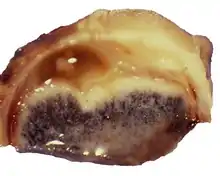
Pigmentierter neuroektodermaler Tumor
Der Pigmentierte neuroektodermale Tumor ist ein sehr seltener Tumor des Schädels bei Kindern. Meist tritt er im Oberkiefer bei Kleinkindern auf. Er gilt als lokal aggressiv, ansonsten als gutartig.[1][2][3]
| Klassifikation nach ICD-10 | |
|---|---|
| D36.9 | Gutartige Neubildung an nicht näher bezeichneter Lokalisation |
| ICD-10 online (WHO-Version 2019) | |
| Klassifikation nach ICD-O-3 | |
|---|---|
| 9363/0 | Pigmentierter neuroektodermaler Tumor Melanoameloblastom Melanotisches Progonom Retinalanlage-Tumor |
| ICD-O-3 erste Revision online | |

Der Tumor entspringt der Neuralleiste und gehört zu den Neuroektodermalen Tumoren.[4]
Synonyme sind: Melanoameloblastom; (veraltet: Melanotisches Progonom; Retinalanlage-Tumor[2]); englisch Melanotic neuroectodermal tumor of infancy; MNTI
Die Erstbeschreibung stammt aus dem Jahre 1918 als „Kongenitales Melanocarcinom“ durch E. Krompecher.[5](zit. Nach[6])
Die Bezeichnung „Melanotic Neuroectodermal Tumor of Infancy“ wurde im Jahre 1966 von E. D. Borello und R. J. Gorlin vorgeschlagen.[7]
Verbreitung
Die Häufigkeit ist nicht bekannt, seit der Erstbeschreibung wurde über etwa 500 Betroffene berichtet. Fast alle Patienten sind bei Entdeckung des Tumors unter einem Jahr alt, etwa 80 % unter 6 Monaten. Ob eines der Geschlechter häufiger betroffen ist, wird in der Literatur uneinheitlich beantwortet.[4][8][6]
Klinische Erscheinungen
Klinische Kriterien sind:[6][2]
- Manifestation im Alter von wenigen Monaten
- Lokalisation in mehr als 2/3 im Oberkiefer
- Schnell wachsende Raumforderung, nur manchmal offensichtlich Melanine enthaltend, mitunter Zahnverlagerung oder Verformung des Mundbereichtes mit Fütterungsproblemen
Der Tumor gilt als gutartig, aber lokal aggressiv und kann maligne entarten.
Diagnose
Die Diagnose ergibt sich mithilfe der medizinische Bildgebung[6] und wird immunhistochemisch bestätigt.[1] Der Tumor produziert Vanillinmandelsäure,[7] aber nur in 35 % mit hohem Plasmaspiegel.[4]
Therapie
Die bevorzugte Behandlung ist die vollständige operative Entfernung.[1][6] Bei etwa 15 % kommt ein Wiederauftreten innerhalb der ersten 12 Monate vor. Als Adjuvante Therapie kommt eine Chemotherapie infrage.[6][2]
Literatur
- A. Kamgarpour, N. Riaz-Montazer u. a.: Melanotic Progonoma: An Unusual Pathology for an Infantile Midline Scalp Mass. In: Pediatric Neurosurgery, 49, 2014, S. 254, doi:10.1159/000363192.
- S. Hamilton, D. Macrae, S. Agrawal, D. Matic: Melanotic neuroectodermal tumour of infancy. In: The Canadian journal of plastic surgery = Journal canadien de chirurgie plastique, Band 16, Nummer 1, 2008, S. 41–44, doi:10.1177/229255030801600108, PMID 19554164, PMC 2690629 (freier Volltext).
- P. Agarwal, S. Saxena, S. Kumar, R. Gupta: Melanotic neuroectodermal tumor of infancy: Presentation of a case affecting the maxilla. In: Journal of oral and maxillofacial pathology: JOMFP, Band 14, Nummer 1, Januar 2010, S. 29–32, doi:10.4103/0973-029X.64309, PMID 21180456, PMC 2995998 (freier Volltext).
- L. A. Renner, A. E. Abdulai: Melanotic neuroectodermal tumour of infancy (progonoma) treated by radical maxillary surgery. In: Ghana medical journal. Band 43, Nummer 2, Juni 2009, S. 90–92, doi:10.4314/gmj.v43i2.55322, PMID 21326849, PMC 3039233 (freier Volltext)
- D. J. Fowler, J. Chisholm, D. Roebuck, L. Newman, M. Malone, N. J. Sebire: Melanotic neuroectodermal tumor of infancy: clinical, radiological, and pathological features. In: Fetal and pediatric pathology. Band 25, Nummer 2, 2006 Mar-Apr, S. 59–72, doi:10.1080/15513810600788715, PMID 16908456.
Einzelnachweise
- M. Guschmann, H. Tönnies, H. Neitzel, G. Schmidt, M. von Wickede, B. Stöver: Kongenitaler melanotischer neuroektodermaler Tumor. In: Monatsschrift Kinderheilkunde, 153, 2005, S. 157, doi:10.1007/s00112-003-0861-4.
- V. Shankar: Pigmented neuroectodermal tumor of infancy. Pathology Outlines
- emedicine
- M. H. Almomani, R. M. Rentea: Melanotic Neuroectodermal Tumor Of Infancy. In: StatPearls [Internet], 2020; ncbi.nlm.nih.gov
- E. Krompecher: Zur Histogenese und Morphologie der Adimantinome und sonstiger Kiefergeschwülste. In: Beitr Path Anat., Band 64, S. 165–197, 1918
- Radiopaedia
- E. D. Borello, R. J. Gorlin: Melanotic neuroectodermal tumor of infancy--a neoplasm of neural crese origin. Report of a case associated with high urinary excretion of vanilmandelic acid. In: Cancer, 1966, Band 19, Heft 2, S. 196–206, PMID 285905, doi:10.1002/1097-0142(196602)19:2<196::aid-cncr2820190210>3.0.co;2-6.
- H. Selim, S. Shaheen, K. Barakat, A. A. Selim: Melanotic neuroectodermal tumor of infancy: review of literature and case report. In: Journal of pediatric surgery, Band 43, Nummer 6, Juni 2008, S. E25–E29, doi:10.1016/j.jpedsurg.2008.02.068, PMID 18558161 (Review).