Bändermodell (Proteine)
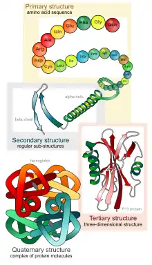
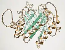
Bändermodelle sind Molekülmodelle von Proteinstrukturen.[1][2][3][4] Sie visualisieren Elemente der Sekundärstruktur wie eine α-Helix oder ein β-Faltblatt und erlauben eine Darstellung der Tertiärstruktur. Verschiedene Polypeptide können zur besseren Unterscheidung farblich unterschiedlich markiert sein. Bändermodelle werden bei der molekularen Modellierung eingesetzt,[5][6] z. B. beim Protein-Engineering. Das Bändermodell wurde 1980/81 durch Jane Richardson etabliert.[1][2][7] Ähnliche Ansätze gab es aber schon früher.
Strukturdarstellungen
| Sekundärstruktur[1][2] | |
|---|---|
| α-Helices | Zylindrische spiralförmige Bänder, mit der Bänderebene ungefähr auf der Peptidbindungsebene. |
| β-Stränge | Pfeile mit einer Dicke von 25 % der Breite in Richtung von N- zu C-Terminus. β-Faltblätter bestehen aus parallel angeordneten β-Strängen mit β-Schleifen. |
| Schleifen und andere | |
| Nichtrepetitive Schleifen | Runde Stränge entlang der α-C-Atome der Aminosäuren, die vom Vordergrund zum Hintergrund dünner werden. |
| Verbindungen zwischen Schleifen und Helices | Runde Stränge entlang der α-C-Atome der Aminosäuren, die beim Übergang zur α-Helix flacher werden. |
| Andere Merkmale | |
| N- und C-Terminus | Kleine Pfeile an einem oder beiden Enden oder mit Buchstaben codiert. Für β-Stränge ist die Richtung des Pfeils ausreichend. Oftmals wird hierfür ein Farbgradient entlang der Aminosäuresequenz verwendet. |
| Disulfidbrücken | Mit verschränktem Doppel-S oder mit einem Zick-Zack-Strich |
| Prosthetische Gruppen oder Inhibitoren | Stabmodell, Kugel-und-Stabmodell |
| Metalle | Kugeln |
| Schattierung und Farbe | Der Kontrast nimmt zum Hintergrund hin ab |
Software
Verschiedene Programme zur Darstellung von Proteinen im Bändermodell wurden entwickelt, z. B. Molscript von Arthur M. Lesk, Karl Hardman und John Priestle,[8] Jmol,[9] DeepView,[10] MolMol,[11] KiNG,[12] UCSF Chimera[13] und PyMOL von Warren DeLano.[14]
Literatur
Einzelnachweise
- Jane Shelby Richardson: The anatomy and taxonomy of protein structure. In: Advances in protein chemistry. Band 34, 1981, S. 167–339, PMID 7020376.
- J. S. Richardson: Schematic drawings of protein structures. In: Methods in enzymology. Band 115, 1985, S. 359–380, PMID 3853075.
- J. S. Richardson: Early ribbon drawings of proteins. In: Nature Structural Biology. Band 7, Nummer 8, August 2000, S. 624–625, doi:10.1038/77912. PMID 10932243. PDF (Memento des Originals vom 1. Februar 2014 im Internet Archive) Info: Der Archivlink wurde automatisch eingesetzt und noch nicht geprüft. Bitte prüfe Original- und Archivlink gemäß Anleitung und entferne dann diesen Hinweis..
- M. Morange: What history tells us XXV. Construction of the ribbon model of proteins (1981). The contribution of Jane Richardson. In: Journal of Biosciences. Band 36, Nummer 4, September 2011, S. 571–574, PMID 21857104. PDF.
- P. D. Sun, C. E. Foster, J. C. Boyington: Overview of protein structural and functional folds. In: Current protocols in protein science / editorial board, John E. Coligan ... [et al.]. Chapter 17 Mai 2004, S. Unit 17.1, doi:10.1002/0471140864.ps1701s35. PMID 18429251.
- Mike Carson, Charles E Bugg: Algorithm for ribbon models of proteins. In: Journal of Molecular Graphics. 4, 1986, S. 121–122, doi:10.1016/0263-7855(86)80010-8.
- J. S. Richardson: Early ribbon drawings of proteins. In: Nature structural biology. Band 7, Nummer 8, August 2000, S. 624–625, doi:10.1038/77912. PMID 10932243. PDF (Memento des Originals vom 1. Februar 2014 im Internet Archive) Info: Der Archivlink wurde automatisch eingesetzt und noch nicht geprüft. Bitte prüfe Original- und Archivlink gemäß Anleitung und entferne dann diesen Hinweis..
- P. J. Kraulis: MOLSCRIPT: a program to produce both detailed and schematic plots of protein structures. In: Journal of Applied Crystallography. 24, S. 946, doi:10.1107/S0021889891004399.
- A. Herráez: Biomolecules in the computer: Jmol to the rescue. In: Biochemistry and molecular biology education : a bimonthly publication of the International Union of Biochemistry and Molecular Biology. Band 34, Nummer 4, Juli 2006, S. 255–261, doi:10.1002/bmb.2006.494034042644, PMID 21638687.
- M. U. Johansson, V. Zoete, O. Michielin, N. Guex: Defining and searching for structural motifs using DeepView/Swiss-PdbViewer. In: BMC Bioinformatics. Band 13, 2012, S. 173, doi:10.1186/1471-2105-13-173, PMID 22823337, PMC 3436773 (freier Volltext).
- R. Koradi, M. Billeter, K. Wüthrich: MOLMOL: a program for display and analysis of macromolecular structures. In: Journal of molecular graphics. Band 14, Nummer 1, Februar 1996, S. 51–5, 29, PMID 8744573.
- V. B. Chen, I. W. Davis, D. C. Richardson: KING (Kinemage, Next Generation): a versatile interactive molecular and scientific visualization program. In: Protein science : a publication of the Protein Society. Band 18, Nummer 11, November 2009, S. 2403–2409, doi:10.1002/pro.250, PMID 19768809, PMC 2788294 (freier Volltext).
- T. D. Goddard, C. C. Huang, T. E. Ferrin: Software extensions to UCSF chimera for interactive visualization of large molecular assemblies. In: Structure (London, England : 1993). Band 13, Nummer 3, März 2005, S. 473–482, doi:10.1016/j.str.2005.01.006, PMID 15766548.
- A. T. Brunger, J. A. Wells: Warren L. DeLano 21 June 1972-3 November 2009. In: Nature structural & molecular biology. Band 16, Nummer 12, Dezember 2009, S. 1202–1203, doi:10.1038/nsmb1209-1202, PMID 19956203.