Mesosom
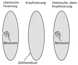
Mesosomen sind Einstülpungen der Plasmamembran von Bakterien, die vermutlich durch chemische Fixierung erzeugt werden. Sie wurden erstmals 1953 bei elektronenmikroskopischen Präparaten beobachtet.[1] Verschiedene Funktionen wurden daraufhin in den 1960er Jahren vorgeschlagen, bis sich seit Ende der 1970er Jahre herausstellte, dass es sich vermutlich um Artefakte handelt.
Beobachtet wurden Mesosomen an Gram-positiven Bakterien.[3] Der Name geht auf eine Arbeit aus dem Jahr 1959 zurück.[4] Eine Funktion der Mesosomen wurde als Organellen bei verschiedenen zellulären Vorgängen vermutet, so etwa die für den Bau der Zellwand während der Zellteilung, Verdopplung des bakteriellen Chromosoms oder als Ort der Oxidativen Phosphorylierung.[5][6]
Ende der 1970er Jahre sammelten sich Daten an, die darauf schließen ließen, dass es sich bei Mesosomen um Strukturen handelt, die erst durch chemische Fixierung hervorgerufen werden, da sie nicht bei Zellen auftraten, die mit anderen Methoden fixiert wurden.[3][7][8] Mit dem Fortschritt in elektronenmikroskopischen Fixierungsmethoden (Kryofixierung und ‚freeze substitution‘) stellten mehrere Arbeiten fest, dass Mesosomen nicht in lebenden Zellen vorkommen.[9][10][11] Einige Forscher waren jedoch der Ansicht, dass die Datenlage nicht schlüssig sei und dass Mesosomen möglicherweise nicht in allen Fällen Artefakte seien.[12][13]
Im Jahr 2000 wurden Mesosomen-ähnliche Einstülpungen an Bakterien beobachtet, die antibakteriellen Peptiden, sogenannten Defensinen ausgesetzt wurden,[14] im Jahr 2007 an solchen, die mit bestimmten Antibiotika behandelt wurden.[15] Das Erscheinungsbild dieser Mesosomen-ähnlichen Strukturen ist möglicherweise eine Folge der Schädigung der Plasmamembran oder der Zellwand durch diese Chemikalien.[16]
Die „Entdeckung“ der Mesosomen und ihre spätere Entlarvung als Artefakt diente in der Wissenschaftstheorie als praktisches Beispiel für den Prozess der Falsifikation einer wissenschaftlichen Idee oder Hypothese. An diesem Beispiel konnte man studieren, wie die Wissenschaftsgemeinde (englisch: ‚Scientific Community‘) dieses Testverfahren durchgeführt und die ursprüngliche Hypothese schließlich zurückgewiesen hat.[17][18][1]
Einzelnachweise
- D. Allchin: The Epistemology of Error. In: Philosophy of Science Association Meetings Vancouver, November. 2000.
- N. Nanninga: The mesosome of Bacillus subtilis as affected by chemical and physical fixation. In: J. Cell Biol. Band 48, Nr. 1, 1971, S. 219–224, PMID 4993484.
- M. T. Silva, J. C. Sousa, J. J. Polónia, M. A. Macedo, A. M. Parente: Bacterial mesosomes. Real structures or artifacts? In: Biochim. Biophys. Acta. Band 443, Nr. 1, 1976, S. 92–105, PMID 821538.
- J. D. Robertson: The ultra structure of cell membranes and their derivatives, Biochem. In: Soc. Syrup. 1959, S. 3.
- A. Suganuma: Studies on the fine structure of Staphylococcus aureus. In: J Electron Microsc (Tokyo). Band 15, Nr. 4, 1966, S. 257–261, PMID 5984369.
- R. D. Pontefract, G. Bergeron, F. S. Thatcher: Mesosomes in Escherichia coli. In: J. Bacteriol. Band 97, Nr. 1, 1969, S. 367–375, PMID 4884819.
- H. R. Ebersold, J. L. Cordier, P. Lüthy: Bacterial mesosomes: method dependent artifacts. In: Arch. Microbiol. Band 130, Nr. 1, 1981, S. 19–22, PMID 6796029.
- M. L. Higgins, H. C. Tsien, L. Daneo-Moore: Organization of mesosomes in fixed and unfixed cells. In: J. Bacteriol. Band 127, Nr. 3, 1976, S. 1519–1523, PMID 821934.
- A. Ryter: Contribution of new cryomethods to a better knowledge of bacterial anatomy. In: Ann. Inst. Pasteur Microbiol. Band 139, Nr. 1, 1988, S. 33–44, PMID 3289587.
- N. Nanninga, G. J. Brakenhoff, M. Meijer, C. L. Woldringh: Bacterial anatomy in retrospect and prospect. In: Antonie Van Leeuwenhoek. Band 50, Nr. 5–6, 1984, S. 433–460, PMID 6442119.
- J. Dubochet, A. W. McDowall, B. Menge, E. N. Schmid, K. G. Lickfeld: Electron microscopy of frozen-hydrated bacteria. In: J. Bacteriol. Band 155, Nr. 1, 1983, S. 381–390, PMID 6408064.
- John F. Stolz: Structure of Phototrophic Prokaryotes. CRC Press, 1991, ISBN 0-8493-4814-5. (books.google.com)
- K. Murata, S. Kawai, B. Mikami, W. Hashimoto: Superchannel of Bacteria: Biological Significance and New Horizons. In: Bioscience, Biotechnology, and Biochemistry. 2008, S. 801080710 (Online).
- C. L. Friedrich, D. Moyles, T. J. Beveridge, R. E. Hancock: Antibacterial action of structurally diverse cationic peptides on gram-positive bacteria. In: Antimicrob. Agents Chemother. Band 44, Nr. 8, 2000, S. 2086–2092, PMID 10898680.
- L. Santhana Raj, H. L. Hing, O. Baharudin u. a.: Mesosomes are a definite event in antibiotic-treated Staphylococcus aureus ATCC 25923. In: Trop Biomed. Band 24, Nr. 1, 2007, S. 105–109, PMID 17568383.
- D. L. Balkwill, S. E. Stevens: Effects of penicillin G on mesosome-like structures in Agmenellum quadruplicatum. In: Antimicrob. Agents Chemother. Band 17, Nr. 3, 1980, S. 506–509, PMID 6775592.
- S. Culp: Defending Robustness: The Bacterial Mesosome as a Test Case. In: PSA: Proceedings of the Biennial Meeting of the Philosophy of Science Association. Band 1994, 1994, S. 46–57, JSTOR:193010.
- N. Rasmussen: Evolving Scientific Epistemologies and the Artifacts of Empirical Philosophy of Science: A Reply Concerning Mesosomes. In: Biology and Philosophy. Band 16, Nr. 5, 2001, S. 627–652 (Online [PDF]).