Makropinozytose
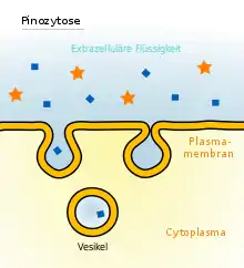
Als Makropinozytose (auch Pinozytose) bezeichnet man die Aufnahme von Flüssigkeitsmengen und darin gelösten Substanzen aus dem Umgebungsmedium einer Zelle in ihr Inneres. Die Aufnahme in das Zellplasma erfolgt in Form von Vesikeln von maximal 150 nm Durchmesser.[1] Das Vesikel wird als Makropinosom bezeichnet.
Das Wort leitet sich aus dem griechischen 'pinein' = trinken und 'kytos' = Hülle ab.[2]
Eigenschaften
Die Makropinozytose ist neben der Phagozytose, der Clathrin-vermittelten Endozytose und der Caveolae-vermittelten Endozytose ein Mechanismus der Endozytose. Sie kommt unter anderem in Phagozyten vor.[3] Die Flüssigkeit wird unspezifisch ohne Beteiligung von Rezeptoren für den Inhalt aufgenommen.[4] Die Vesikel verschmelzen anschließend mit Endosomen und Lysosomen im Endomembransystem. Im Verlauf der Bildung von Makropinosomen wird Rab7 vermehrt gebildet. Die IP3-Kinase und die Phospholipase C sind an der Bildung beteiligt, cAMP führt dagegen zu einer Wiedereingliederung des Vesikels in die Zellmembran. Während der Bildung von Makropinosomen werden die Phosphoinositide PI(4,5)P2 und PI(3,4,5)P3 gebildet und die Rho-GTPase inaktiviert.[5] Der Transport der Makropinosomen erfolgt entlang des Aktin-Zytoskeletts.[5]
Zelleintritt für Pathogene
Das Ebolavirus, das Vacciniavirus, Adenoviren, manche Picornaviren und das humane Herpesvirus 8 verwenden Makropinosom-ähnliche Vesikel für den Zelleintritt.[6][7][8] Das von EHEC gebildete Vero-Toxin, sowie Salmonellen, Legionella pneumophila und einige Protozoen werden über Makropinosomen in Zellen aufgenommen.[9][10]
Weblinks
- IUPAC Gold Book, "pinocytosis". Abgerufen am 3. Juli 2016.
Einzelnachweise
- Daniel Boujard, Bruno Anselme, Christophe Cullin, Céline Raguénès-Nicol: Zell- und Molekularbiologie im Überblick. Springer Spektrum, Berlin, Heidelberg 2014, ISBN 978-3-642-41761-0, S. 226, doi:10.1007/978-3-642-41761-0.
- http://www.biologie-schule.de/pinozytose.php
- R. Levin, S. Grinstein, D. Schlam: Phosphoinositides in phagocytosis and macropinocytosis. In: Biochimica et Biophysica Acta. Band 1851, Nummer 6, Juni 2015, S. 805–823, doi:10.1016/j.bbalip.2014.09.005, PMID 25238964.
- J. P. Lim, P. A. Gleeson: Macropinocytosis: an endocytic pathway for internalising large gulps. In: Immunology and cell biology. Band 89, Nummer 8, November 2011, S. 836–843, doi:10.1038/icb.2011.20, PMID 21423264.
- Y. Egami, T. Taguchi, M. Maekawa, H. Arai, N. Araki: Small GTPases and phosphoinositides in the regulatory mechanisms of macropinosome formation and maturation. In: Frontiers in physiology. Band 5, 2014, S. 374, doi:10.3389/fphys.2014.00374, PMID 25324782, PMC 4179697 (freier Volltext).
- J. Mercer, A. Helenius: Virus entry by macropinocytosis. In: Nature cell biology. Band 11, Nummer 5, Mai 2009, S. 510–520, doi:10.1038/ncb0509-510, PMID 19404330.
- M. F. Saeed, A. A. Kolokoltsov, T. Albrecht, R. A. Davey: Cellular entry of ebola virus involves uptake by a macropinocytosis-like mechanism and subsequent trafficking through early and late endosomes. In: PLoS pathogens. Band 6, Nummer 9, 2010, S. e1001110, doi:10.1371/journal.ppat.1001110, PMID 20862315, PMC 2940741 (freier Volltext).
- M. Valiya Veettil, S. Sadagopan, N. Kerur, S. Chakraborty, B. Chandran: Interaction of c-Cbl with myosin IIA regulates Bleb associated macropinocytosis of Kaposi's sarcoma-associated herpesvirus. In: PLoS pathogens. Band 6, Nummer 12, 2010, S. e1001238, doi:10.1371/journal.ppat.1001238, PMID 21203488, PMC 3009604 (freier Volltext).
- M. C. Kerr, J. T. Wang, N. A. Castro, N. A. Hamilton, L. Town, D. L. Brown, F. A. Meunier, N. F. Brown, J. L. Stow, R. D. Teasdale: Inhibition of the PtdIns(5) kinase PIKfyve disrupts intracellular replication of Salmonella. In: The EMBO journal. Band 29, Nummer 8, April 2010, S. 1331–1347, doi:10.1038/emboj.2010.28, PMID 20300065, PMC 2868569 (freier Volltext).
- T. M. de Carvalho, E. S. Barrias, W. de Souza: Macropinocytosis: a pathway to protozoan infection. In: Frontiers in physiology. Band 6, 2015, S. 106, doi:10.3389/fphys.2015.00106, PMID 25914647, PMC 4391238 (freier Volltext).