HBcAg
HBcAg (Hepatitis B core antigen) ist ein Protein des Hepatitis-B-Virus (HBV).
| HBcAg | ||
|---|---|---|
| Andere Namen |
Core antigen, Capsid protein | |
| Masse/Länge Primärstruktur | 185 Aminosäuren, 21.332 Da | |
| Bezeichner | ||
| Externe IDs | ||
| Orthologe (Hepatitis-B-Virus) | ||
| Entrez | 944568 | |
| UniProt | P03148
| |
| PubMed-Suche | 944568 | |
Eigenschaften

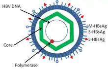
HBcAg ist das Kapsidprotein des HBV. Es bildet Multimere bis hin zu einem ikosaedrischen Kapsid aus 240 HBcAg mit einer Triangulationszahl T = 4 und 180 HBcAg mit T = 3.[1] Nach Entpackung wird das HBcAg in der Zelle entlang Mikrotubuli in den Zellkern transportiert.[1] Eine Phosphorylierung des HBcAg exponiert eine Signalsequenz nahe dem C-Terminus (Position 187–204) zum Import in den Zellkern über Importin α und β.[1] Im Zellkern bindet das Kapsid an NUP153.[1] Dort erfolgt die Entpackung der HBV-Genome, sofern sie vollständig gereift sind.[1] Allerdings werden verschiedene unvollständige virale und virusartige Partikel gebildet.[2]
An der Position 1 bis 144 ist die Kapsiddomäne, während ab 150 bis 185 eine Arginin-reiche, Protamin-artige Nukleinsäure-bindende Domäne liegt.[3]
HBeAg wird aus dem gleichen offenen Leseraster wie HBcAg gebildet. Neugebildetes HBcAg im Zytosol bindet prä-genomische RNA (an der HBcAg-Position 206–212) und die Polymerase Protein P, woraufhin das DNA-Genom des HBV anhand der RNA-Vorlage erzeugt wird.[1] Der N-Terminus enthält vier Cysteine, die vermutlich einen Zinkfinger bilden.[4] Anschließend erfolgt entweder ein erneuter Import in den Zellkern oder das Kapsid bindet an das endoplasmatische Retikulum zur Knospung.[1]
Tapasin bindet an den Abschnitt 18–27 des HBcAg und verstärkt die Aktivität von zytotoxischen T-Zellen gegen HBV.[5]
Anwendungen
HBcAg wird zum Nachweis einer momentanen Infektion verwendet. Nach einmaliger Infektion bleibt das HBc-Antigen während der Infektion immer positiv. Hierbei kann man eine gegen HBV geimpfte Person von einer zum jetzigen Zeitpunkt oder schon früher erkrankten Person mit HBV unterscheiden.[6][7]
Ebenso wird es zur Behandlung von Hepatitis-B-Infektionen als Target untersucht.[8]
Literatur
- B. Böttcher, S. A. Wynne, R. A. Crowther: Determination of the fold of the core protein of hepatitis B virus by electron cryomicroscopy. In: Nature. Band 386, Nummer 6620, März 1997, S. 88–91, doi:10.1038/386088a0, PMID 9052786.
- A. Zlotnick, B. Venkatakrishnan, Z. Tan, E. Lewellyn, W. Turner, S. Francis: Core protein: A pleiotropic keystone in the HBV lifecycle. In: Antiviral research. Band 121, September 2015, S. 82–93, doi:10.1016/j.antiviral.2015.06.020, PMID 26129969, PMC 4537649 (freier Volltext).
Einzelnachweise
- Uniprot: C - Capsid protein - Hepatitis B virus genotype A2 subtype adw2 (strain Rutter 1979) (HBV-A) - C gene & protein. In: uniprot.org. 20. Juni 2018, abgerufen am 26. Februar 2018 (englisch).
- J. Hu, K. Liu: Complete and Incomplete Hepatitis B Virus Particles: Formation, Function, and Application. In: Viruses. Band 9, Nummer 3, 03 2017, S. , doi:10.3390/v9030056, PMID 28335554, PMC 5371811 (freier Volltext).
- M. Seifer, D. N. Standring: A protease-sensitive hinge linking the two domains of the hepatitis B virus core protein is exposed on the viral capsid surface. In: Journal of virology. Band 68, Nummer 9, September 1994, S. 5548–5555, PMID 7520091, PMC 236955 (freier Volltext).
- Pfam: pfam08290 Hep_core_N: Hepatitis core protein, putative zinc finger.
- X. Chen, Y. Tang, Y. Zhang, M. Zhuo, Z. Tang, Y. Yu, G. Zang: Tapasin modification on the intracellular epitope HBcAg18-27 enhances HBV-specific CTL immune response and inhibits hepatitis B virus replication in vivo. In: Laboratory investigation; a journal of technical methods and pathology. Band 94, Nummer 5, Mai 2014, S. 478–490, doi:10.1038/labinvest.2014.6, PMID 24614195.
- T. Kimura, A. Rokuhara, A. Matsumoto, S. Yagi, E. Tanaka, K. Kiyosawa, N. Maki: New enzyme immunoassay for detection of hepatitis B virus core antigen (HBcAg) and relation between levels of HBcAg and HBV DNA. In: Journal of clinical microbiology. Band 41, Nummer 5, Mai 2003, S. 1901–1906. ISSN 0095-1137, PMID 12734224, PMC 154683 (freier Volltext).
- T. Cao, P. Meuleman, I. Desombere, M. Sällberg, G. Leroux-Roels: In vivo inhibition of anti-hepatitis B virus core antigen (HBcAg) immunoglobulin G production by HBcAg-specific CD4(+) Th1-type T-cell clones in a hu-PBL-NOD/SCID mouse model. In: Journal of virology. Band 75, Nummer 23, Dezember 2001, S. 11449–11456. ISSN 0022-538X, doi:10.1128/JVI.75.23.11449-11456.2001, PMID 11689626, PMC 114731 (freier Volltext).
- L. Y. Mak, D. K. Wong, W. K. Seto, C. L. Lai, M. F. Yuen: Hepatitis B core protein as a therapeutic target. In: Expert opinion on therapeutic targets. Band 21, Nummer 12, 12 2017, S. 1153–1159, doi:10.1080/14728222.2017.1397134, PMID 29065733.