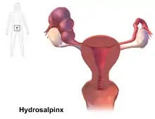
Hydrosalpinx
Die Hydrosalpinx, von altgriechisch ὕδωρ hydor, deutsch ‚Wasser‘ und altgriechisch σαλπίγξ salpinx, deutsch ‚Eileiter‘, lateinisch Sactosalpinx serosa, ist eine mit Flüssigkeit gefüllte Aufweitung des Eileiters durch Aufstau wässrigen Sekretes meist nach einer abgelaufenen Salpingitis.[1][2][3]
| Klassifikation nach ICD-10 | |
|---|---|
| N70.1 | Chronische Salpingitis und Oophoritis – einschl. Hydrosalpinx |
| ICD-10 online (WHO-Version 2019) | |
Ist die Flüssigkeit eitrig, spricht man von einer Pyosalpinx, enthält sie Blut, liegt eine Hämatosalpinx vor.[4]
.jpg.webp)
Ursache
Als Ursache für den Verschluss der Tube am distalen Ende, der Eileiterampulle, oder an beiden Enden kommen infrage:[2]
- Unterleibsentzündung mit Ausbildung von Verklebungen (häufigste Ursache) nach Salpingitis (Entzündung des Eileiters), Adnexitis, Oopheritis (Entzündung des Eierstocks), Endometritis
- Endometriose, oft mit einer Hämatosalpinx
- Hysterektomie
- Tubenligatur
- bösartige Tumoren der Tube
- Ovulationsinduktion[5]
Klinische Erscheinungen
Eine Hydrosalpinx kann klinisch unauffällig sein oder mit Unterleibsbeschwerden einhergehen, nicht selten kann auch eine Unfruchtbarkeit bestehen.[6][2]
Es besteht eine Assoziation mit erheblich verminderter Erfolgsrate nach künstlicher Befruchtung, die Ursache ist noch nicht geklärt.[7][3]
Diagnose
Die Diagnose kann mit Hilfe verschiedener bildgebender Verfahren gestellt werden: mit der Hystero-Kontrast-Salpingographie, mittels Computertomographie, heute mittels Magnetresonanztomographie oder als Methode der Wahl durch die Sonographie.[2][8][3][9]
Differentialdiagnose
Abzugrenzen sind:[2]
- Zysten neben dem Eierstock
- zystische Neubildungen des Eierstockes
- erweiterte Darmschlingen
- erweiterte Beckenvenen
- Tarlov-Zyste im Becken
Therapie
Eine Behandlung ist bei Beschwerden sowie vor künstlicher Befruchtung angezeigt.[10] Infrage kommen die laparoskopische Entfernung des Eileiters (Salpingektomie), die Salpingostomie oder andere Eingriffe.[11][12]
Komplikationen
Als Komplikation kann es zu einer Infektion der Flüssigkeit mit Ausbildung einer Pyosalpinx kommen.[13]
Geschichte
Die Erstbeschreibung stammt wohl von Reinier de Graaf.[14][15]
Literatur
- L. M. Della Grotta, R. B. Dyer, B. L. Holbert: The "cogwheel" sign of hydrosalpinx. In: Abdominal radiology, Band 44, Nummer 10, 10 2019, S. 3486–3487, doi:10.1007/s00261-019-02144-7, PMID 31346740 (Review).
- H. M. Harb, J. Ghosh, F. Al-Rshoud, B. Karunakaran, I. D. Gallos, A. Coomarasamy: Hydrosalpinx and pregnancy loss: a systematic review and meta-analysis. In: Reproductive biomedicine online, Band 38, Nummer 3, März 2019, S. 427–441, doi:10.1016/j.rbmo.2018.12.020, PMID 30665848.
Einzelnachweise
- Eintrag zu Hydrosalpinx im Flexikon, einem Wiki der Firma DocCheck
- Radiopaedia
- Emedicine Fallopian Tube Disorders
- Willibald Pschyrembel: Klinisches Wörterbuch. 266., aktualisierte Auflage. de Gruyter, Berlin 2014, ISBN 978-3-11-033997-0
- V. L. Schiller, K. Tsuchiyama: Development of hydrosalpinx during ovulation induction. In: Journal of Ultrasound in Medicine. Band 14, Nummer 11, November 1995, S. 799–803, doi:10.7863/jum.1995.14.11.799, PMID 8551543.
- K. von Horn, A. Doster, A. Freis, S. Hecht, T. Strowitzki: Hydrosalpinx. In: Gynäkologische Endokrinologie. 15, 2017, S. 260, doi:10.1007/s10304-017-0139-x.
- A. N. Andersen, Z. Yue, F. J. Meng, K. Petersen: Low implantation rate after in-vitro fertilization in patients with hydrosalpinges diagnosed by ultrasonography. In: Human reproduction. Band 9, Nummer 10, Oktober 1994, S. 1935–1938, doi:10.1093/oxfordjournals.humrep.a138362, PMID 7844229.
- Y. Zalel, D. Soriano, S. Lipitz, S. Mashiach, R. Achiron: Contribution of color Doppler flow to the ultrasonographic diagnosis of tubal abnormalities. In: Journal of Ultrasound in Medicine. Band 19, Nummer 9, September 2000, S. 645–649, doi:10.7863/jum.2000.19.9.645, PMID 10972562.
- O. Benjaminov, M. Atri: Sonography of the abnormal fallopian tube. In: American Journal of Roentgenology. Band 183, Nummer 3, September 2004, S. 737–742, doi:10.2214/ajr.183.3.1830737, PMID 15333364.
- A. Volodarsky-Perel, W. Buckett, T. Tulandi: Treatment of hydrosalpinx in relation to IVF outcome: a systematic review and meta-analysis. In: Reproductive biomedicine online. Band 39, Nummer 3, September 2019, S. 413–432, doi:10.1016/j.rbmo.2019.04.012, PMID 31324437 (Review).
- N. P. Johnson, W. Mak, M. C. Sowter: Surgical treatment for tubal disease in women due to undergo in vitro fertilisation. In: The Cochrane database of systematic reviews. Nummer 3, 2004, S. CD002125, doi:10.1002/14651858.CD002125.pub2, PMID 15266464 (Review).
- K. Y. Ng, Y. Cheong: Hydrosalpinx – Salpingostomy, salpingectomy or tubal occlusion. In: Best practice & research. Clinical obstetrics & gynaecology. Band 59, August 2019, S. 41–47, doi:10.1016/j.bpobgyn.2019.01.011, PMID 30824209 (Review).
- B. Nikolic, K. Nguyen, L. G. Martin, D. C. Redd, I. Best, M. I. Silverstein: Pyosalpinx developing from a preexisting hydrosalpinx after uterine artery embolization. In: Journal of vascular and interventional radiology: JVIR. Band 15, Nummer 3, März 2004, S. 297–301, doi:10.1097/01.rvi.0000116187.30591.00, PMID 15028817.
- W. M. Ankum, H. L. Houtzager, O. P. Bleker: Reinier De Graaf (1641–1673) and the fallopian tube. In: Human reproduction update. Band 2, Nummer 4, 1996 Jul-Aug, S. 365–369, doi:10.1093/humupd/2.4.365, PMID 9080233.
- B. I. Tshisuaka: Graaf, Reinier de. In: Werner E. Gerabek, Bernhard D. Haage, Gundolf Keil, Wolfgang Wegner (Hrsg.): Enzyklopädie Medizingeschichte. De Gruyter, Berlin / New York 2005, ISBN 3-11-015714-4, S. 506.