B-DNA
B-DNA ist eine der möglichen Doppelhelix-Strukturen von DNA.[1]
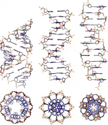
Seitenansicht und Obenansicht von A-, B- und Z-DNA
Eigenschaften
B-DNA ist eine rechtsgängige doppelsträngige DNA-Doppelhelix, die im Vergleich zur A-Form entspannter ist und deren Nukleinbasen orthogonal zur Helixachse ausgerichtet sind. B-DNA ist die unter physiologischen Bedingungen typisch vorliegende Form doppelsträngiger DNA, z. B. bei Chromosomen in Zellen.[2] Andere DNA-Strukturen sind z. B. A-DNA, Z-DNA, hairpin, triplex, cruciform, left-handed Z-form, tetraplex und A-motif.[3] Aufgrund der entspannteren Form besitzt B-DNA im Vergleich zu A-DNA eine kleinere Anzahl an Nukleinbasen pro Windung der Helix, eine flachere große Furche, eine tiefere kleine Furche und ist um etwa 30 % länger pro Windung.[4]
Doppelsträngige DNA-Strukturen (dsDNA)
B-DNA

Basenpaargeometrien
| Geometrie | A-Form | B-Form | Z-Form |
|---|---|---|---|
| Helix-Drehrichtung | rechtsgängig | rechtsgängig | linksgängig |
| Wiederholungseinheit | 1 bp | 1 bp | 2 bp |
| Rotation/bp | 33,6° | 34,6° | 60°/2 |
| bp/Windung | 11 | 10,4 | 12 |
| Inklination der bp zur Achse | +19° | −1,2° | −9° |
| Länge/bp entlang der Achse | 2,4 Å (0,24 nm) | 3,4 Å (0,34 nm) | 3,7 Å (0,37 nm) |
| Länge/Windung | 24,6 Å (2,46 nm) | 33,2 Å (3,32 nm) | 45,6 Å (4,56 nm) |
| Biegung (propeller twist) | +18° | +16° | 0° |
| Glycosylwinkel | anti | anti | Pyrimidin: anti, Purin: syn |
| Phosphatabstand | 5,9 Å | 7,0 Å | C: 5,7 Å, G: 6,1 Å |
| Glycosylflexibilität (sugar pucker) | C3’-endo | C2'-endo | C: C2'-endo, G: C3’-endo |
| Durchmesser | 23 Å (2,3 nm) | 20 Å (2,0 nm) | 18 Å (1,8 nm) |
Literatur
- E. Girard, T. Prangé, A. C. Dhaussy, E. Migianu-Griffoni, M. Lecouvey, J. C. Chervin, M. Mezouar, R. Kahn, R. Fourme: Adaptation of the base-paired double-helix molecular architecture to extreme pressure. In: Nucleic acids research. Band 35, Nummer 14, 2007, S. 4800–4808, doi:10.1093/nar/gkm511, PMID 17617642, PMC 1950552 (freier Volltext).
- P. Cysewski: The post-SCF quantum chemistry characteristics of inter- and intra-strand stacking interactions in d(CpG) and d(GpC) steps found in B-DNA, A-DNA and Z-DNA crystals. In: Journal of molecular modeling. Band 15, Nummer 6, Juni 2009, S. 597–606, doi:10.1007/s00894-008-0378-9, PMID 19039609.
Weblinks
- Nucleic Acid Nomenclature (englisch).
Einzelnachweise
- D. L. Beveridge, T. E. Cheatham, M. Mezei: The ABCs of molecular dynamics simulations on B-DNA, circa 2012. In: Journal of biosciences. Band 37, Nummer 3, Juli 2012, S. 379–397, PMID 22750978, PMC 4029509 (freier Volltext).
- A. S. Boyer, S. Grgurevic, C. Cazaux, J. S. Hoffmann: The human specialized DNA polymerases and non-B DNA: vital relationships to preserve genome integrity. In: Journal of molecular biology. Band 425, Nummer 23, November 2013, S. 4767–4781, doi:10.1016/j.jmb.2013.09.022, PMID 24095858.
- J. Choi, T. Majima: Conformational changes of non-B DNA. In: Chemical Society reviews. Band 40, Nummer 12, Dezember 2011, S. 5893–5909, doi:10.1039/c1cs15153c, PMID 21901191.
- M. C. Wahl, M. Sundaralingam: Crystal structures of A-DNA duplexes. In: Biopolymers. Band 44, Nummer 1, 1997, S. 45–63, PMID 9097733, doi:10.1002/(SICI)1097-0282(1997)44:1<45::AID-BIP4>3.0.CO;2-#.
This article is issued from Wikipedia. The text is licensed under Creative Commons - Attribution - Sharealike. The authors of the article are listed here. Additional terms may apply for the media files, click on images to show image meta data.